Online available: 2025-09-10
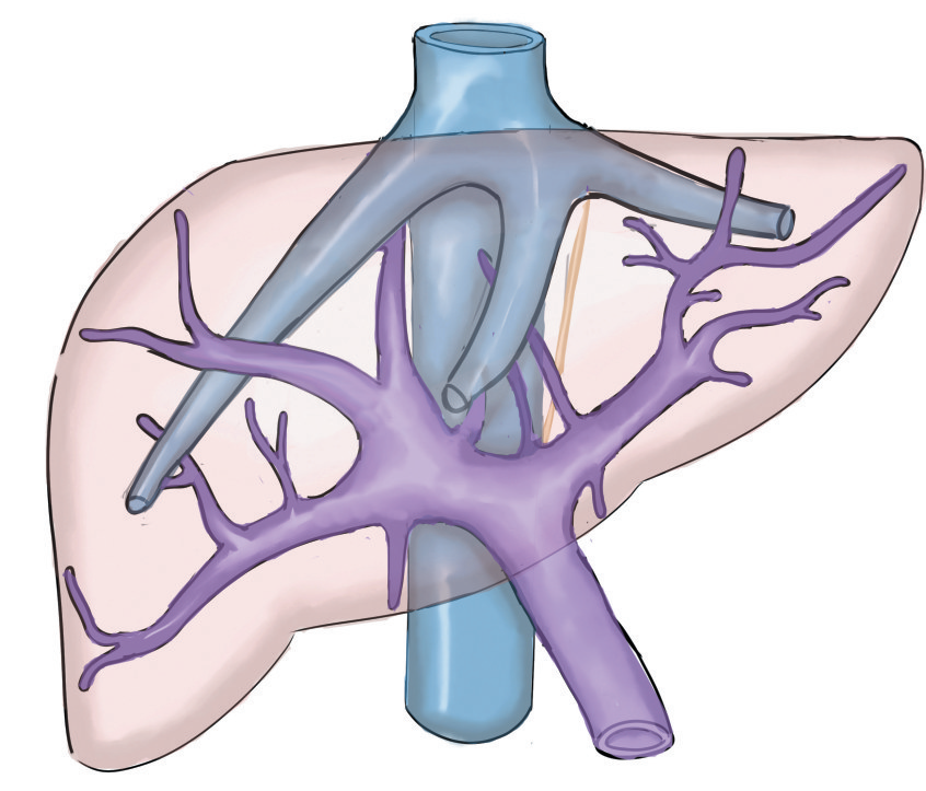
In recent years, with the development of high-resolution imaging, three-dimensional visualization, indocyanine green fluorescence imaging, virtual reality, and augmented reality, liver anatomy has advanced from traditional macroscopic morphological description to a multi-dimensional era of individualized, microscopic, and functional fine anatomy. The latest progress in liver fine anatomy and its innovative role in surgical strategies include: (1) the evolution from Couinaud’s segmentation to individualized watershed resection based on sub-segments; (2) the re-recognition of intrahepatic anatomical spaces; (3) the transition from static anatomy to dynamic watersheds, the development of two-step hepatectomy with combined liver partition and portal vein ligation, and hepatic vein deprivation, which utilize shunt vessels to pre-regulate the watershed and preserve liver parenchyma; (4) the integration of function and morphology, the assessment of regional liver function through SPECT, MRI hepatobiliary-specific contrast agents, and other techniques; (5) the precise demarcation of tumor invasion boundaries by molecular imaging technology to reduce the positive margin rate in non-anatomical resection; (6) the promotion of fine anatomy and surgical innovation by artificial intelligence (AI). In the future, with the in-depth research of AI, molecular imaging, and hemodynamics, liver surgery will further develop towards precision, functionality, and intelligence, providing safer and more effective individualized treatment plans for patients with complex liver tumors.