Only by attaching importance to the study of clinical applied anatomy, we can grasp the essence of gynecological surgery and the operation can be safe. Anatomically,gynecologic and obstetric operations centered on the cervical region are the types of operations that we must master,such as various types of Q-M classification operation for cervical cancer,DIE operation around the cervix,complex cesarean section,pop operation,etc. Therefore,the anatomy of the three-dimensional ring around the cervix is the clinical anatomical basis for obstetrics and gynecology surgery. This article focuses on the introduction of the three-dimensional ring around the cervix,a new clinical anatomical concept named by our according to the needs of surgery. Only by fully understanding and thoroughly mastering the anatomy in the three-dimensional ring around the cervix and the spatial three-dimensional relationship between each anatomical structure,we can complete the most basic surgery in obstetrics and gynecology.
Vesicovaginal fistula is a disease that seriously affects women's physical and mental health and quality of life. Repair of vesicovaginal fistula is an important means of treatment for this disease. Because it conforms to the concept of minimally invasive surgery, has small trauma,quick recovery,no scar,few complications,is easy for patients to accept,can be repeated after failure,and has a short hospital stay,and low medical expenses,it is widely used by gynecologists. The anatomical key points of this operation are: to find the original fistula of the vesicovaginal fistula;to remove the scar around the fistula;to fully free the tissue around the fistula,to suture without tension and to have healthy fistula tissue and good blood supply;to use the centrifugal repair method combined with the centripetal repair method to carry out multi-layer suture of the fistula.
Radical hysterectomy is the primary treatment for patients with early-stage cervical cancer. The Querleu-Morrow classification utilizes fixed anatomical structures as landmarks to promote a more precise resection range. A thorough understanding of the critical anatomical structures, such as the paracervical spaces,ligaments, vessels, nerves, and the ureteral tunnel, is the basis for adequate resection and safe surgery.
Para-aortic lymphadenectomy is an important part of the surgical treatment for gynecological malignancies,which is of great significance for the staging and treatment of malignant tumors. Aortaventralis accompanies postcava,and is adjacent to the kidney,ureter and intestine,so the surgical field is difficult to expose. Para-aortic lymphadenectomy is difficult and has many complications,which calls for higher requirements in anatomical knowledge and operation skills of the operator. This paper expounds the anatomical problems that need attention in performing this operation.
Pelvic lymph node dissection is an important part of the evaluation and management of the staging and prognosis of gynecological malignant tumors. Its success is directly related to the patient's survival rate and quality of life. It is one of the necessary skills that gynecological oncologists need to master. Whether it can be completed in a standardized and safe manner depends largely on the doctor's in-depth understanding of the pelvic anatomical structure. This article explores the anatomical issues related to pelvic lymph node dissection to provide a reference for clinical surgery.
The presacral area is situated between the posterior wall of the rectum and the sacrum,encompassing loose connective tissue,the middle sacral artery,lateral sacral blood vessels,the presacral venous plexus,presacral nerves,and presacral lymph nodes. It is essential to comprehend the direction and distribution of arteries and veins within the presacral area,the anatomy of the sacral nerves and their influence on visceral function,as well as the relationship between the anterior sacral foramen,sacral vertebrae,and the vascular network. Furthermore,based on findings from traditional autopsy and CTA three-dimensional reconstructions of the presacral vascular area, it is proposed to delineate the surgical safe zone for sacral fixation. A thorough understanding of the anatomical positioning and interrelations of these structures offers an anatomical foundation for presacral nerve resection,presacral lymphadenectomy,and other surgical procedures,thereby providing both a theoretical basis and practical guidance for presacral surgery.
Endometrial cancer surgery is the preferred minimally invasive surgery in gynecological tumors. Under the tumor-free principle,the popularization of laparoscopic radical resection of endometrial cancer represents the progress in diagnosis and treatment technology. In recent years,the promotion and application of surgical robots represented by Da Vinci has impacted the importance of traditional laparoscopy to a certain extent. Some scholars call it “advanced laparoscopy”,which represents the recognition of this technology. Based on the interpretation of the characteristics of surgical robots and the key operations of endometrial cancer surgery,we think about the differences between the two procedures from the anatomical level,and explore how to achieve the most complete tumor resection,minimal adjacent structural and functional damage and minimal traumatic stress to the body,so as to truly achieve “bloodless surgical field and accurate dissection”.
Total laparoscopic hysterectomy is a common gynecological surgery,which has been favored by patients and doctors in recent years. Although total laparoscopic hysterectomy carries a low risk of ureteral injury,it can have serious consequences. The identification of the high risk factors of ureteral injury and the anatomical features of the high risk factors can reduce the occurrence of ureteral injury. If necessary,the ureteral stent can be used for anatomical identification to complete the operation.
Placental accreta spectrum disorders (PAS) seriously threatens the physical health of pregnant women. This article explores the high-risk factors for PAS,including cesarean section,endometrial injury and repair abnormalities,especially cesarean scar pregnancy; It elucidates the pathological mechanism of PAS,vascular remodeling caused by vascular endothelial growth factor (VEGF),and abnormal invasion of placental villus tissue into the uterine muscle layer; The article highlights the regional anatomical classification method of PAS and its application in clinical surgery as well as the key anatomical features and surgical methods of different surgical procedures in PAS cesarean section surgery.
Postpartum hemorrhage can be managed effectively by surgical suture. Vascular ligation,compared to tissue suture techniques,is a surgical procedure that is challenging for novices to master and has a relatively higher incidence of complications. Vascular anatomy serves as a crucial theoretical foundation for vascular ligation. This article expounds on the vascular anatomy related to vascular ligation surgery,the anatomical differences between the gestational and non-gestational periods,surgical skills,and the prevention and management of complications.
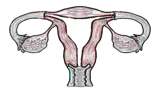
Adnexectomy is a common and basic surgical procedure in the field of gynecology. Nonetheless,it is necessary to consider some critical anatomical issues during the procedure,including the potential risk of injury to major blood vessels and the ureters. The purpose of this article is to provide a detailed analysis of the surgical procedure and associated techniques,based on the anatomical structure of the pelvic cavity.